Keloid vs Hypertrophic Scar: Key Differences, Symptoms, and Management Approaches
- Sep 20, 2025
- 12 min read
Updated: Feb 25
A raised scar can be unsettling — but not all raised scars are the same. Two of the most commonly confused types are keloid scars and hypertrophic scars. While both result from the skin producing excess collagen during wound healing, they behave quite differently: one tends to stay within the original wound boundaries and may improve over time, while the other can spread beyond the wound and is less likely to resolve on its own.
Understanding this distinction matters because the management approach for each is different. This article explains what sets them apart — in terms of appearance, growth pattern, symptoms, and the options a doctor may discuss with you — based on current clinical understanding.
How Raised Scars Form
When skin is injured — by surgery, a burn, a deep cut, severe acne, or even a piercing — the body responds by producing collagen to close and repair the wound. In most cases, this results in a flat, faded scar that gradually becomes less noticeable over months to years.
In some people, however, the collagen-producing cells (fibroblasts) become overactive, generating more fibrous tissue than is needed. This imbalance between collagen synthesis and collagen breakdown can lead to a raised, thickened scar. Depending on how far that excess tissue spreads, the result is either a hypertrophic scar or a keloid. Both are benign (non-cancerous) and pose no direct health threat, but they can cause discomfort and cosmetic concern — and they require different management strategies.
What Is a Hypertrophic Scar?
A hypertrophic scar is a raised, firm scar that develops within the boundaries of the original wound. It arises from an exaggerated but spatially contained healing response — the body over-produces collagen at the injury site, but that excess tissue does not invade the surrounding, uninjured skin.
Appearance and Characteristics
Hypertrophic scars are typically raised and firm to the touch, and may feel rubbery. They are usually red or pink in the early months and tend to soften and become paler as they mature. Crucially, the scar remains within the edges of the original wound — this is the single most important clinical feature that distinguishes it from a keloid.
Texture: Firm, raised, sometimes slightly ridged
Colour: Red or pink initially; may fade to flesh-tone over time
Boundaries: Confined to the original wound margins
Timeline: Usually appears within weeks of the injury; may evolve over several months
Natural course: Has a tendency to partially or fully flatten over one to two years in many cases, though this varies by individual and scar severity
Common Causes and Risk Factors
Hypertrophic scarring tends to develop when there is significant tension, prolonged inflammation, or delayed healing at the wound site. Areas of the body where the skin is under constant mechanical stress — the chest, shoulders, and joints — are particularly prone.
Surgical incisions — especially on the chest, shoulders, and upper back where skin tension is high
Burns — second- and third-degree burns frequently lead to hypertrophic scarring due to the prolonged inflammatory phase
Traumatic wounds — deep lacerations, abrasions, and puncture wounds
Inflammatory acne — particularly nodular or cystic acne lesions that rupture or are manipulated
Wound infections or delayed healing — prolonged inflammation increases the risk of abnormal collagen deposition
Body piercings — especially in cartilaginous areas such as the ear helix
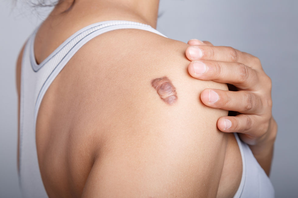
What Is a Keloid Scar?
A keloid is a type of pathological scar characterised by the growth of fibrous tissue that extends beyond the original wound boundaries into surrounding, previously uninjured skin. This invasive growth pattern is the defining feature of a keloid and sets it apart from all other scar types.
Keloids are considered a disorder of wound healing rather than simply an aggressive form of scarring. The underlying biology involves overactive wound healing signals that cause fibroblasts to continue producing collagen long after the wound has closed — unlike a normal scar, keloid tissue lacks the self-limiting mechanism that stops growth, which is why keloids can continue to enlarge over months to years.
Defining Features of Keloids
Extension beyond wound margins: The scar spreads into surrounding normal skin — this is the hallmark sign
Surface texture: Typically shiny, smooth, and dome-shaped, though some have an irregular, lobulated form
Colour: Ranges from pink or red in lighter skin tones to dark brown or purple in deeper skin tones
Growth pattern: May continue to grow slowly for months or years; spontaneous regression is uncommon
Symptoms: Itching, tenderness, a burning or prickling sensation — often more pronounced during the active growth phase
Who Is at Higher Risk?
Keloid formation has a clear genetic component. Research has shown that individuals of African, Asian, and Hispanic descent have a statistically higher prevalence of keloid formation compared to those of European descent, though keloids can occur in any skin type. A positive family history significantly increases an individual's risk.
Common high-risk sites include the earlobes (particularly after piercing), the chest (especially the sternum), the upper back, and the shoulders. The type of triggering injury also matters: burns, chickenpox scars, severe inflammatory acne, and surgical wounds in high-tension areas carry a higher risk of keloid formation in susceptible individuals. In highly susceptible people, even very minor trauma — including insect bites or superficial abrasions — can occasionally trigger a keloid.
Key Differences at a Glance
The table below summarises the main clinical distinctions between hypertrophic scars and keloids. These differences guide diagnosis and influence which management approaches a doctor is likely to recommend.
Feature | Hypertrophic Scar | Keloid Scar |
Boundary | Remains within original wound margins | Extends beyond original wound into surrounding skin |
Onset after injury | Weeks (typically within 4–8 weeks) | Weeks to months (sometimes over 1 year later) |
Growth pattern | May stabilise and partially regress over time | May continue to grow slowly; rarely self-resolves |
Spontaneous improvement | More common; many soften and flatten over time | Uncommon without intervention |
Colour | Red/pink early; may fade over time | Pink, red, dark brown, or purple depending on skin tone |
Texture | Firm, raised, may feel rubbery | Firm, shiny, smooth or lobulated surface |
Symptoms | Mild itching or tenderness; generally improves as scar matures | Itching, burning, tenderness — may persist during growth phase |
Common locations | Any high-tension area (joints, chest, shoulders); follows wound shape | Earlobes, sternum, shoulders, upper back; may be disproportionate to wound size |
Genetic predisposition | Less prominent | Strong; higher prevalence in darker skin tones and in those with family history |
Recurrence risk after surgery | Lower (though possible) | Higher; recurrence is a recognised clinical challenge |
Management Approaches: Hypertrophic Scars
Because hypertrophic scars have a natural tendency to improve over time, management often begins with conservative, non-invasive measures. The goal is to support the scar's maturation, reduce symptoms, and improve its appearance. The most appropriate approach depends on the scar's size, location, age, and how it is affecting the patient.
Silicone-Based Products
Silicone gel sheeting and silicone gel formulations are among the most studied non-invasive options for hypertrophic scar management. Clinical guidelines have long recognised silicone-based products as a first-line, non-invasive option for the prevention and management of hypertrophic scars — a position supported by multiple published reviews in this area. The proposed mechanism involves sustained hydration of the outer skin layer (stratum corneum), which may help moderate fibroblast activity and collagen synthesis, though the precise mechanism remains an area of ongoing research.
Silicone products are available over the counter and are typically applied to clean, dry scar tissue for extended periods each day, with consistent use over several months generally recommended. They are well-tolerated across skin types and ages.
Pressure Therapy
Continuous mechanical pressure applied to a healing scar — via custom-fitted compression garments, elastic bandages, or specialised dressings — is a recognised approach particularly for larger hypertrophic scars resulting from burns or extensive surgery. The mechanical force is thought to reduce local blood flow and influence collagen fibre organisation, potentially limiting excessive scar thickening. Pressure therapy requires consistent, long-term use (often many hours per day over months) and is most effective when initiated early in the scar maturation process.
Corticosteroid Injections
For persistent or symptomatic hypertrophic scars that have not responded adequately to conservative measures, corticosteroid injections directly into the scar tissue (intralesional injections) are a well-established clinical option. A corticosteroid medication — triamcinolone acetonide, a type of anti-inflammatory steroid — is injected by a doctor into the scar. Its anti-inflammatory properties may help moderate fibroblast activity and collagen synthesis, potentially reducing scar volume and alleviating symptoms such as itching and discomfort.
Treatment is typically delivered in a series of sessions, commonly spaced four to six weeks apart. Potential side effects — including thinning of the skin (atrophy), skin lightening (hypopigmentation), and small visible blood vessels (telangiectasia) — are well-documented and should be discussed with the treating doctor before commencing treatment. These risks are particularly relevant for patients with darker skin tones.
Laser Therapy
Energy-based devices can be considered for hypertrophic scars when other measures have not achieved the desired result. Vascular lasers such as the Vbeam (pulsed dye laser, 595 nm) target abnormal blood vessels within the scar, which may help reduce redness and improve scar texture. Ablative fractional laser devices, including fractional CO2 lasers, can be used to remodel scar tissue by creating controlled micro-injuries that stimulate collagen reorganisation. The choice of device, treatment settings, and number of sessions are determined by the treating doctor based on each patient's scar characteristics and skin type.
Management Approaches: Keloid Scars
Keloids are generally more challenging to manage than hypertrophic scars and often require a combination of therapies. Treating with a single modality alone is associated with higher recurrence rates for keloids, particularly surgical excision when used without additional treatment. A doctor will assess the keloid's size, location, age, the patient's skin type, and their personal and family history before recommending a management plan.
A note on keloid management: Treating keloids requires balancing the potential benefit of each approach against the risk of triggering further keloid formation from the treatment itself — for example, from a surgical incision. This is why treatment planning for keloids is highly individualised and requires careful medical judgement.
Corticosteroid Injections
Corticosteroid injections directly into the keloid tissue are frequently used as a foundational treatment, either as a standalone measure for smaller or early-stage keloids, or in combination with other approaches. As with hypertrophic scars, a corticosteroid medication (triamcinolone acetonide) is injected by a doctor, and a course of repeat sessions over several months is typically needed. Potential side effects including skin thinning, skin lightening, and small visible blood vessels are possible and should be discussed with the treating doctor.
Cryotherapy
Cryotherapy involves the controlled application of extreme cold — most commonly liquid nitrogen — to keloid tissue, administered by a doctor in clinic. The cold is thought to disrupt the cellular structure of the keloid and its blood supply, potentially reducing its size. Cryotherapy is generally most suitable for smaller keloids and is commonly combined with corticosteroid injections rather than used alone. Multiple sessions are typically required. Temporary skin lightening (hypopigmentation) is a recognised side effect, which is an important consideration for patients with darker skin tones.
Surgical Excision with Adjunctive Therapy
Surgical removal of keloid tissue can significantly reduce its bulk and improve its appearance. However, surgical excision alone carries a well-documented risk of keloid recurrence — in some cases, the resulting surgical wound itself may trigger a new or larger keloid. For this reason, surgery for keloids is almost always combined with additional treatments to reduce recurrence risk, which may include:
Corticosteroid injections into the wound, commenced at or shortly after surgery and continued post-operatively
Silicone gel sheeting applied to the healing surgical wound
Radiotherapy — typically initiated within 24–48 hours of surgery for higher-risk keloids
The decision to proceed with surgery, and which additional therapies to combine, is made by the treating doctor in discussion with the patient, taking into account the keloid's characteristics and the individual's risk profile.
Laser Therapy
Laser treatments may be considered as part of a combined keloid management plan. The Vbeam pulsed dye laser may help reduce redness and improve the vascularity of keloid tissue, and some published studies suggest it may also reduce symptoms such as itching when used over multiple sessions. Ablative fractional CO2 lasers may be considered to address surface texture. The evidence base for laser therapy in keloids continues to evolve, and outcomes vary between individuals. Treatment decisions should be made in consultation with a doctor experienced in scar management.
Radiotherapy
Post-operative radiotherapy is a recognised adjunct to surgical excision for keloids, particularly for larger, recurrent, or surgically complex cases. It is typically administered in controlled, low doses by a qualified professional, beginning as soon as possible after surgery — usually within 24 to 48 hours. The rationale is that radiation may help limit the activity of fibroblasts in the healing wound, potentially reducing the likelihood of keloid reformation.
Prevention for High-Risk Individuals
For individuals with a personal or family history of keloids, proactive wound care and lifestyle modifications may help reduce — though not entirely eliminate — the risk of abnormal scarring after future skin injuries or procedures. These measures are best discussed with a doctor in the context of each person's risk profile.
Optimise Wound Care from the Start
The quality of wound care in the days and weeks following an injury can influence how the scar develops. Key principles include keeping the wound clean with gentle daily cleansing, maintaining a moist wound environment with petroleum jelly or a recommended wound care product, and protecting the area with a sterile, non-adherent dressing. Infection prolongs inflammation and increases the risk of abnormal scarring, so prompt attention to any signs of wound infection is important.
In areas of high skin tension — such as the chest, shoulders, and over joints — minimising mechanical stress on the healing wound where possible may be beneficial. Surgical wounds in these areas may be managed with tension-offloading dressings or taping techniques as advised by the treating doctor.
Avoid Elective Skin Procedures in High-Risk Areas
Individuals who are known to form keloids are generally advised to carefully weigh the risks of elective procedures that break the skin — including cosmetic tattooing, body piercings, and non-essential procedures — particularly on the earlobes, chest, shoulders, and upper back. Before any planned procedure, discussing your scarring history with the treating doctor allows appropriate precautions to be taken.
Use Proactive Scar Management Measures Early
Once a wound has fully closed, starting scar management measures early may help influence how the scar matures. Silicone gel or silicone sheeting applied consistently over several months is one of the most widely recommended measures for individuals at elevated risk. Pressure dressings or garments may be recommended for larger surgical wounds or burns.
New scars are also sensitive to UV exposure, which can cause lasting discoloration. Protecting healing scars with a broad-spectrum sunscreen (SPF 30 or higher) and physical coverage is advisable until the scar has fully matured.
When to Consult a Doctor
Raised scars that are troublesome, uncertain in diagnosis, growing, or causing functional limitation warrant professional assessment. A doctor or skin specialist can accurately identify the type of scar, rule out other skin conditions, and discuss a management plan appropriate for the individual. Consider seeking a medical evaluation if:
You are unsure whether your raised scar is a hypertrophic scar or a keloid
The scar continues to grow or spread beyond its original boundaries months after the injury
The scar is causing persistent itching, burning, tenderness, or discomfort
The scar is located over a joint and may be affecting your range of motion
The scar's appearance is causing distress and you wish to explore management options
You have a personal or family history of keloids and have sustained a new skin injury or are planning a procedure
There are signs of wound infection such as increasing redness, warmth, swelling, or discharge
Consulting a doctor early — particularly when there is a family history of keloid formation — may allow for more proactive planning for current and future wound care.
Frequently Asked Questions
Can a hypertrophic scar turn into a keloid?
A true hypertrophic scar does not convert into a keloid, as they are considered distinct conditions with different underlying biology. However, some scars that appear initially confined to the wound may in fact be early keloids that have not yet spread — which is one reason that professional evaluation is helpful when there is diagnostic uncertainty.
Do keloids go away on their own?
Spontaneous regression of keloids without treatment is uncommon. While some keloids may become less symptomatic over many years, significant self-resolution without intervention is not reliably expected. Hypertrophic scars, by contrast, have a greater tendency to flatten and fade over time.
Are keloids dangerous?
Keloids are benign (non-cancerous) growths and do not pose a direct health risk. However, they can cause significant physical discomfort — including itching, tenderness, and pain — as well as psychological distress, and may affect movement if located over joints.
Is there a way to confirm whether I have a keloid?
A clinical diagnosis is usually made by an experienced doctor based on the scar's history, appearance, and growth pattern. In cases of diagnostic uncertainty, a skin biopsy can be performed to examine the tissue under a microscope, as the structural features of keloids differ from those of hypertrophic scars. Your doctor will advise whether a biopsy is appropriate in your case.
Is darker skin more prone to keloids?
Epidemiological data consistently shows a higher prevalence of keloid formation in individuals of African, Asian, and Hispanic heritage compared to those of European descent. However, keloids can and do occur in all skin types, and having a darker skin tone does not mean an individual will necessarily develop keloids — individual genetic predisposition remains an important factor.
Can keloids come back after treatment?
Keloid recurrence after treatment is a well-recognised clinical challenge, particularly after surgical excision performed without additional therapy. Combined approaches — such as surgery followed promptly by radiotherapy and/or corticosteroid injections — are associated with lower recurrence rates in published studies, though recurrence cannot be entirely excluded in any individual case. Long-term follow-up with a doctor is often recommended.
Summary
Hypertrophic scars and keloids are both raised scars caused by excess collagen production during wound healing, but they behave differently and require different management approaches.
A hypertrophic scar stays within the original wound boundaries, often improves on its own over time, and typically responds well to conservative measures such as silicone products, pressure therapy, or corticosteroid injections. A keloid, by contrast, grows beyond the wound margin, is unlikely to resolve without intervention, and often requires a combination of treatments — such as corticosteroid injections, surgical excision with adjunctive therapy, or laser treatments — to manage effectively and reduce the risk of recurrence.
For individuals with darker skin tones or a family history of keloids, the likelihood of developing a keloid after a skin injury is higher, and early consultation with a doctor can help guide preventive care. Regardless of scar type, professional medical evaluation is the most reliable way to confirm the diagnosis and identify the most appropriate management plan for each individual.
Please see below for treatment details.
Note: This article is for educational purposes only and does not constitute medical advice. Individual treatment plans should be developed in consultation with qualified healthcare professionals. Treatment outcomes vary from person to person, and no guarantee of results is intended or implied. All professional treatments mentioned should be performed by licensed medical practitioners in Singapore, using HSA-approved products, devices, and techniques, as applicable.


