PIE vs PIH: Complete Guide to Post-Inflammatory Skin Discoloration and Treatment Options
- Nov 10, 2025
- 12 min read
Updated: Nov 14, 2025
When inflammation subsides, many individuals notice persistent marks on their skin. These discolorations, known as post-inflammatory erythema (PIE) and post-inflammatory hyperpigmentation (PIH), can significantly impact self-confidence. Understanding the distinction between these conditions is essential for selecting appropriate treatment approaches.
Quick Answer: What's the Main Difference?
The fundamental difference between PIE and PIH lies in their underlying cause:
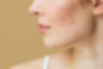
PIE (Post-Inflammatory Erythema) appears as red or pink marks caused by dilated blood vessels beneath the skin surface. This vascular condition results from inflammation-induced changes to capillaries.
PIH (Post-Inflammatory Hyperpigmentation) manifests as brown, gray, or dark patches due to excess melanin production triggered by skin inflammation. This pigmentary condition is particularly common in individuals with darker skin tones.
Understanding Post-Inflammatory Erythema (PIE)
What Is PIE?
Post-inflammatory erythema represents a vascular response to skin inflammation. When inflammation occurs, blood vessels in the affected area may dilate and proliferate. Even after the initial inflammation resolves, these vascular changes can persist, creating visible redness that characterizes PIE.
What Causes PIE?
PIE can develop following various inflammatory skin conditions and injuries. Common triggers include:
Inflammatory Skin Conditions:
Acne vulgaris, particularly inflammatory lesions (papules, pustules, nodules)
Rosacea, which frequently causes facial redness and flushing
Eczema and contact dermatitis, especially when scratching damages capillaries
Psoriasis, characterized by red, scaly patches
Procedural Causes:
Chemical peels performed at inappropriate depths
Laser treatments without proper parameters or aftercare
Microdermabrasion when overly aggressive
Other cosmetic procedures with excessive skin trauma
Physical Trauma:
Burns (thermal, chemical, or sun-related)
Cuts, scrapes, and abrasions
Insect bites causing localized inflammation
Picking or squeezing blemishes
Clinical Appearance of PIE
PIE typically presents as flat or slightly raised patches ranging from light pink to deep red or purplish tones. The intensity depends on several factors including skin tone, inflammation severity, and the depth of affected blood vessels. Unlike PIH, PIE does not involve melanin changes but rather reflects the presence of dilated capillaries visible through the skin surface. Affected areas may feel slightly warm due to increased blood flow.
Duration of PIE
The persistence of PIE varies considerably among individuals. Mild cases may resolve within several weeks to a few months. More persistent presentations can last six months to over a year. Several factors influence resolution time:
Severity of the initial inflammation
Individual healing capacity
Ongoing inflammation or irritation
Sun exposure and photoprotection practices
Treatment interventions employed
Understanding Post-Inflammatory Hyperpigmentation (PIH)
What Is PIH?
PIH develops when skin inflammation triggers melanocytes (pigment-producing cells) to generate excess melanin. This overproduction causes darkened patches or spots that persist after the inflammatory condition has resolved. PIH represents a pigmentary rather than vascular response to inflammation.
What Causes PIH?
PIH shares many triggering factors with PIE but affects darker skin tones disproportionately due to higher baseline melanocyte activity. Common causes include:
Primary Inflammatory Triggers:
Acne breakouts, particularly when accompanied by inflammation
Eczema flare-ups
Psoriasis lesions
Insect bites and allergic reactions
Mechanical and Chemical Trauma:
Friction or repeated rubbing of the skin
Burns from various sources
Cosmetic procedures including chemical peels, laser treatments, and microdermabrasion when performed without appropriate precautions or aftercare
Risk Factors: Research indicates that individuals with Fitzpatrick skin types III-VI demonstrate increased susceptibility to PIH development. Asian, African, and Latin populations show particularly high prevalence rates, with studies documenting PIH in up to 58% of acne patients in Asian populations.
Clinical Appearance of PIH
PIH manifests as flat, discolored areas on the skin. The color spectrum ranges from light brown to dark brown, gray, or occasionally black, depending on the individual's baseline skin tone and the depth of melanin deposition. These patches appear darker than surrounding unaffected skin and can vary in size from small spots to larger confluent areas.
Types of PIH
Understanding PIH depth is crucial for treatment selection:
Epidermal PIH
Epidermal PIH involves melanin accumulation within the epidermis (outermost skin layer). This type typically appears as light to dark brown macules and responds more favorably to treatment interventions. With appropriate sun protection and topical treatments, epidermal PIH often demonstrates gradual improvement over several months.
Dermal PIH
Dermal PIH occurs when melanin deposits in the dermis (deeper skin layer). This presentation can appear blue-gray, slate gray, or brown and proves more challenging to address. Dermal PIH tends to persist longer than epidermal PIH. Treatment approaches may require more intensive interventions, and consultation with a qualified dermatologist is recommended for optimal management strategies.
Duration of PIH
PIH persistence varies based on multiple factors:
Epidermal PIH: May show improvement within several months with appropriate intervention
Dermal PIH: Can persist for extended periods, sometimes years
Individual variability: Skin type, melanocyte activity, sun exposure, and treatment adherence all influence resolution time
Studies indicate that in Asian populations, PIH persists for at least one year in over 50% of affected individuals, with approximately 22% experiencing discoloration lasting five years or longer.
Key Differences Between PIE and PIH
Appearance Comparison
The visual distinction between PIE and PIH provides the most immediate diagnostic clue:
PIE Characteristics:
Red, pink, or purple coloration
Flat marks that may appear slightly raised
Color intensity can vary with temperature or blood flow
More prominent in fair to medium skin tones
PIH Characteristics:
Brown, gray, or black coloration
Consistently flat macules or patches
Stable color unaffected by temperature
More prevalent in medium to dark skin tones
Underlying Mechanisms
While both conditions arise from inflammation, their pathophysiology differs fundamentally:
PIE Development: Inflammation causes blood vessel dilation and proliferation in the affected area. Even after the inflammatory stimulus resolves, these vascular changes persist. The redness observed in PIE reflects increased erythrocyte concentration in dilated capillaries near the skin surface.
PIH Development: Inflammatory mediators stimulate melanocytes to produce excess melanin. This pigment then deposits in either the epidermis or dermis, creating visible darkening. The inflammation-triggered melanin overproduction continues even after the initial inflammatory condition has subsided.
Duration Patterns
PIE Timeline: Generally demonstrates faster resolution compared to PIH, with most cases improving within several months to one year. However, persistent cases can extend beyond this timeframe.
PIH Timeline: Typically requires longer resolution periods, particularly for dermal PIH. Treatment duration often extends from several months to years, depending on pigment depth and individual factors.
Treatment Approaches for PIE
Post-inflammatory erythema management focuses on reducing redness, addressing vascular changes, and preventing further inflammation. Treatment selection should be individualized based on severity, skin type, and patient preferences. A qualified healthcare professional can provide personalized recommendations.
Vascular Laser and Light-Based Treatments
Laser and light-based technologies represent primary treatment modalities for PIE, targeting the vascular component of the condition.
Pulsed Dye Laser (PDL)
PDL has been extensively studied for vascular lesions and is considered a well-established option for PIE management. Operating at wavelengths around 585-595nm, PDL targets oxyhemoglobin in dilated blood vessels. Research indicates that PDL can provide improvement in PIE appearance, though individual responses vary.
Clinical considerations:
Treatment parameters should be customized to individual skin characteristics
Multiple sessions may be required for optimal outcomes
Individual results vary; not all patients experience the same degree of improvement
Potential side effects include temporary bruising, swelling, and rarely, pigmentation changes
Potassium Titanyl Phosphate (KTP) Laser
KTP lasers operate at 532nm and may be considered for PIE management by qualified practitioners. This wavelength targets superficial blood vessels effectively. Some evidence suggests KTP lasers may also address active inflammatory acne by targeting porphyrins produced by acne-causing bacteria.
Intense Pulsed Light (IPL)
IPL technology utilizes multiple wavelengths to address both vascular and pigmentary concerns. Some studies suggest IPL can be beneficial for PIE, particularly when using vascular filters (typically 560nm or higher). IPL may offer a cost-effective alternative to dedicated vascular lasers, though treatment outcomes can vary among individuals.
Important Note: Laser and light-based treatments should only be performed by licensed medical professionals using HSA-approved devices. Consultation with a qualified practitioner is essential to determine suitability and establish appropriate treatment parameters for your specific condition.
Topical Treatment Options for PIE
While vascular lasers represent primary interventions for PIE, certain topical ingredients may provide supporting benefits in managing redness and inflammation.
Azelaic Acid
Azelaic acid demonstrates anti-inflammatory and antioxidant properties. Research indicates that 15% azelaic acid gel may be beneficial for post-inflammatory erythema management. One study showed improvement in PIE appearance with consistent use over 12 weeks, though individual responses varied.
Niacinamide (Vitamin B3)
Niacinamide strengthens the skin barrier and possesses anti-inflammatory properties. Some evidence suggests it may help reduce redness and improve skin texture. Concentrations of 2-5% are commonly found in skincare formulations.
Vitamin C (L-Ascorbic Acid)
As a potent antioxidant, vitamin C may help reduce inflammation and support collagen synthesis. Some practitioners recommend vitamin C serums as adjunctive therapy for PIE, though high-quality clinical evidence remains limited.
Tranexamic Acid
Emerging research suggests tranexamic acid may have applications in managing post-inflammatory erythema, potentially by reducing vascular endothelial growth and inflammation. However, evidence remains preliminary, and this application should be discussed with a healthcare professional.
Application Guidance: Topical treatments work best when incorporated into a comprehensive skincare routine emphasizing gentle cleansing, consistent moisturization, and diligent sun protection. Results typically require several weeks to months of consistent use.
Treatment Approaches for PIH
Managing post-inflammatory hyperpigmentation requires interventions that reduce melanin production, promote pigment dispersion, and encourage skin cell turnover. Treatment efficacy varies based on PIH type (epidermal vs. dermal), skin tone, and individual factors.
Topical Treatments for PIH
Systematic reviews of PIH treatments indicate that several topical agents demonstrate evidence of efficacy, though individual results vary.
Retinoids
Topical retinoids (including tretinoin, adapalene, and tazarotene) are supported by high-quality evidence for PIH management. Retinoids accelerate skin cell turnover, promoting dispersion of excess melanin. Research indicates retinoids represent one of the most evidence-based topical interventions for PIH, though they require consistent use over several months and may cause initial irritation.
Clinical Considerations:
Start with lower concentrations to assess tolerance
Typically prescribed by healthcare professionals
Sun protection is essential during retinoid use
Improvement generally becomes noticeable after 8-12 weeks of consistent application
Hydroxy Acids
Alpha hydroxy acids (AHAs) such as glycolic acid and beta hydroxy acids (BHAs) like salicylic acid are supported by multiple high-quality studies for PIH management. These agents promote exfoliation and skin cell turnover, helping to fade pigmentation gradually.
Application Notes:
Concentrations vary; higher concentrations should be professionally supervised
May cause mild irritation or sensitivity
Consistent sun protection is crucial
Hydroquinone
Hydroquinone inhibits melanin production and has been studied extensively for hyperpigmentation. In Singapore, hydroquinone products require professional oversight and should only be used under medical supervision. Healthcare professionals can assess suitability and provide appropriate monitoring.
Important Considerations:
Should be used only under medical supervision in Singapore
Treatment duration typically limited to several months
Not suitable for all skin types; professional assessment required
Kojic Acid
Kojic acid inhibits tyrosinase, an enzyme involved in melanin production. Some evidence suggests it may be beneficial for PIH, though research quality varies. Kojic acid is often combined with other depigmenting agents for enhanced effect.
Niacinamide
In addition to its benefits for PIE, niacinamide may help reduce PIH by inhibiting melanosome transfer from melanocytes to keratinocytes. Concentrations of 2-5% are commonly used in over-the-counter formulations.
Vitamin C
L-ascorbic acid and its derivatives may help inhibit melanin production and provide antioxidant protection. Evidence for vitamin C in PIH management is moderate, with some studies showing benefit when combined with other treatments.
Arbutin and Licorice Extract
Plant-derived ingredients like arbutin and licorice extract demonstrate mild melanin-inhibiting properties. While evidence is less robust compared to retinoids or hydroxy acids, these ingredients may provide supporting benefits in PIH management formulations.
Chemical Peels
Chemical peels involve controlled application of chemical solutions to promote exfoliation and skin renewal. Superficial peels using AHAs or salicylic acid may be considered for epidermal PIH management. Deeper peels should only be performed by licensed medical professionals.
Important Considerations:
Peel depth and agent selection must be customized to skin type and PIH characteristics
Inappropriate peel selection or technique can paradoxically worsen PIH
Multiple sessions are typically required
Professional assessment is essential to determine suitability
Microdermabrasion
Microdermabrasion is a minimally invasive procedure that mechanically exfoliates the skin using fine crystals or a diamond-tipped device. It may provide modest improvement in skin texture and mild PIH. However, evidence for its efficacy in PIH is limited compared to other interventions.
Laser Treatments for PIH
Various laser modalities have been investigated for PIH management, with outcomes varying based on laser type, PIH depth, and skin characteristics.
Q-Switched Nd:YAG Laser
Q-switched lasers deliver high-intensity pulses that may help fragment melanin deposits. These treatments are typically reserved for dermal PIH or resistant cases and should only be performed by experienced practitioners using HSA-approved devices.
Clinical Considerations:
Treatment parameters must be carefully selected for darker skin tones to minimize risk
Multiple sessions typically required
Potential complications include temporary darkening or, rarely, worsening of pigmentation
Individual outcomes vary significantly
Fractional Lasers
Fractional laser technologies create controlled micro-injuries to stimulate skin renewal. Some evidence suggests fractional lasers may be beneficial for PIH, though they carry risks of post-inflammatory hyperpigmentation if not appropriately performed, particularly in darker skin tones.
Risk Factors:
Not recommended during active acne breakouts
Requires experienced practitioners for optimal safety
Strict sun protection essential post-treatment
Important Note: According to systematic review evidence published in 2024, laser and energy-based devices achieved complete response in approximately 18% of PIH cases, with varying degrees of improvement in others. Complete resolution cannot be guaranteed, and treatment responses are highly individual. Only licensed medical professionals should perform laser treatments using appropriate, HSA-approved equipment.
Prevention Strategies for PIE and PIH
Preventing post-inflammatory skin discoloration is often more effective than treating established marks. The following strategies may help reduce the risk of developing PIE and PIH:
Sun Protection: The Foundation
Sun exposure represents one of the most significant factors that can worsen both PIE and PIH. UV radiation can darken existing marks and potentially prolong resolution time.
Recommended Sun Protection Practices:
Apply broad-spectrum sunscreen with SPF 30 or higher daily, regardless of weather
Reapply every two hours, especially after swimming or perspiring
Seek shade during peak UV hours (typically 10 AM to 4 PM)
Consider protective clothing including wide-brimmed hats and UV-protective garments
Use physical barriers like umbrellas when prolonged sun exposure is unavoidable
Note for Tropical Climates: Singapore's equatorial location results in consistently high UV index levels throughout the year. Diligent sun protection is particularly important for preventing and managing PIH in this environment.
Avoid Picking, Squeezing, or Trauma
Physical manipulation of inflammatory lesions increases trauma to the skin and surrounding blood vessels, potentially increasing the risk of developing both PIE and PIH.
Protective Behaviors:
Resist the urge to pick or squeeze blemishes
Avoid aggressive rubbing or scrubbing of inflamed areas
Use gentle cleansing techniques
Pat skin dry rather than rubbing vigorously
Consider using hydrocolloid patches for acne lesions to prevent picking
Early Intervention for Inflammatory Conditions
Addressing inflammatory skin conditions promptly may help reduce the severity and duration of subsequent PIE and PIH.
Recommended Approaches:
Consult a healthcare professional for persistent or severe acne
Seek appropriate treatment for eczema, rosacea, psoriasis, and other inflammatory conditions
Follow prescribed treatment regimens consistently
Avoid discontinuing treatments without medical guidance
Report treatment ineffectiveness or side effects to your healthcare provider promptly
Gentle Skincare Practices
Maintaining a gentle, non-irritating skincare routine supports skin barrier function and may help reduce inflammation.
Recommended Practices:
Use mild, fragrance-free cleansers
Avoid harsh physical exfoliants during active inflammation
Introduce new active ingredients gradually
Maintain consistent moisturization to support barrier function
Avoid products with known irritants or allergens for your skin type
Consider Professional Treatment Risks
While cosmetic procedures can address various skin concerns, certain treatments carry risks of triggering PIE or PIH if not appropriately performed.
Risk Mitigation:
Ensure procedures are performed by licensed, experienced practitioners
Discuss your skin type, history of PIH, and concerns before treatment
Follow all pre- and post-treatment instructions carefully
Report unexpected reactions promptly
Allow adequate healing time between treatments
When to Consult a Healthcare Professional
While some cases of PIE and PIH may improve gradually with time and appropriate skincare, certain situations warrant professional medical evaluation:
Indications for Medical Consultation
Persistent or Progressive Discoloration: If PIE or PIH shows no signs of improvement after several months of appropriate self-care, or if discoloration worsens despite intervention, professional assessment is recommended.
Diagnostic Uncertainty: Differentiating PIE from PIH, and both conditions from other skin disorders, can be challenging. If you are uncertain about the nature of your skin discoloration, a healthcare professional can provide definitive diagnosis.
Severe or Extensive Involvement: When discoloration covers large areas, appears particularly severe, or significantly impacts quality of life, professional treatment options may be more appropriate than over-the-counter measures alone.
Development of New Symptoms: If discoloration is accompanied by itching, pain, bleeding, textural changes, or other new symptoms, medical evaluation is important to rule out other conditions.
Concerns About Scarring: If marks appear to be developing into keloids, hypertrophic scars, or atrophic scars, timely professional intervention may help optimize outcomes.
Interest in Prescription Treatments: More intensive interventions including prescription-strength retinoids, hydroquinone (where appropriate), or procedural treatments require professional oversight.
Pre-existing Conditions: Individuals with underlying skin conditions, photosensitivity disorders, or taking photosensitizing medications should consult healthcare professionals before pursuing certain treatments.
What to Expect During Consultation
A qualified healthcare professional will typically:
Conduct a thorough examination of affected areas
Assess skin type and any relevant medical history
Distinguish between PIE, PIH, and other potential conditions
Recommend individualized treatment approaches based on your specific situation
Discuss realistic expectations and potential treatment timelines
Review potential risks and benefits of suggested interventions
Provide guidance on appropriate skincare practices
Summary: Key Takeaways
Understanding the distinction between post-inflammatory erythema (PIE) and post-inflammatory hyperpigmentation (PIH) enables more effective management of these common skin concerns.
Essential Differences:
PIE presents as red, pink, or purple marks caused by vascular changes (dilated blood vessels)
PIH appears as brown, gray, or dark patches resulting from excess melanin production
PIE typically resolves faster than PIH, though timelines vary among individuals
PIE is more visible in fair-medium skin tones, while PIH predominantly affects medium-dark skin tones
Treatment Approaches:
PIE management often involves vascular lasers and anti-inflammatory topical treatments
PIH treatment typically includes retinoids, hydroxy acids, and melanin-inhibiting agents supported by systematic review evidence
Treatment outcomes vary significantly among individuals; no guarantee of results is intended or implied
Professional medical assessment is recommended for determining appropriate treatment strategies
Prevention Strategies:
Consistent sun protection represents the single most important preventive measure for both conditions
Avoiding picking or squeezing inflammatory lesions may reduce risk
Early treatment of underlying inflammatory conditions may help minimize post-inflammatory changes
Gentle skincare practices support skin barrier function and may reduce inflammation
When to Seek Professional Care: Consultation with a qualified healthcare professional is recommended for persistent discoloration, diagnostic uncertainty, severe involvement, development of new symptoms, concerns about scarring, or interest in prescription-strength interventions.
Note: This article is for educational purposes only and does not constitute medical advice. Individual treatment plans should be developed in consultation with qualified healthcare professionals. Treatment outcomes vary from person to person, and no guarantee of results is intended or implied. All professional treatments mentioned should be performed by licensed medical practitioners in Singapore, using HSA-approved products, devices, and techniques, as applicable.